
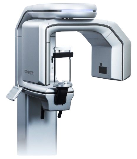
Description
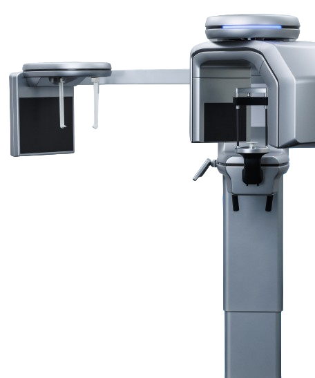
Making more consistent diagnosis and more confident treatment possible with 3D Imaging
PreXion has made high-resolution clinical image quality our first priority in the development of dental cone beam CT. We have over 15 years of experience working with three-dimensional DICOM image processing and software, and we are currently contributing to daily oral pathology diagnostics around the world.
Responsive customer support and strict quality control standards are the defining characteristics of our in-house product development.
Every aspect of the PreXion3D Expedition, from our rigid quality control standards and manufacturing to our comprehensive customer support, is developed in Japan. Our software has been used for more than 15 years in clinical procedures, and We have developed our x-ray generator, a central component of a cone beam CT, in-house.
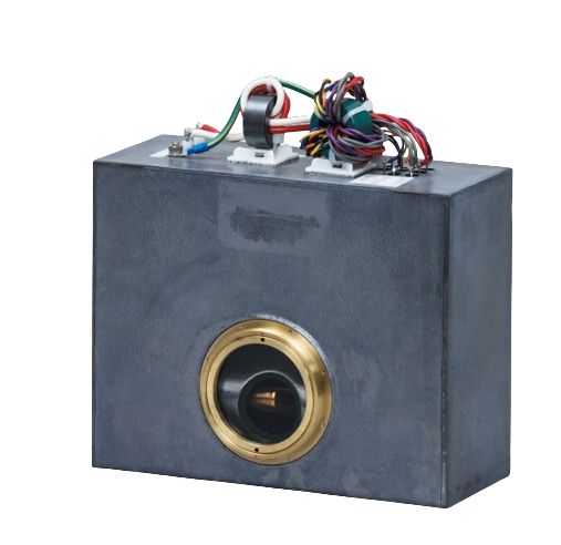
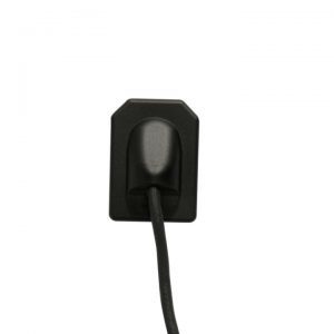
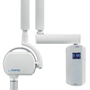
X-ray generator
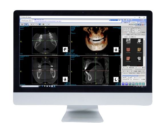
Software
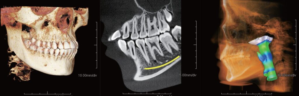
The benefits of incorporating a cone beam CT into daily diagnoses
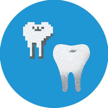
- Practitioners can make diagnoses and provide treatment with more precision by viewing pathology that could not be recognized with conventional 2D images, such as the spatial structure of teeth and jawbone as well as the location of nerves.
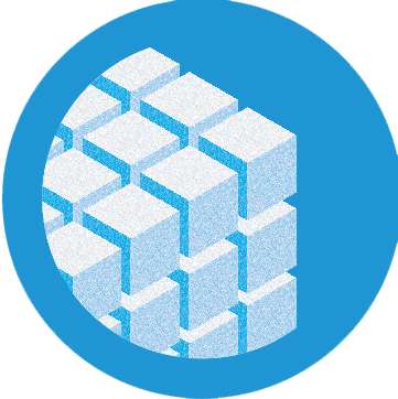
- Cone beam CT allows for low dosage without compromising on precise, clear image quality. This not only ensures accurate diagnosis but also reduces patient concern.
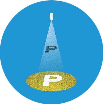
- Easy-to-understand icons and intuitive operation during imaging allow for stress-free diagnosis and better patient understanding.
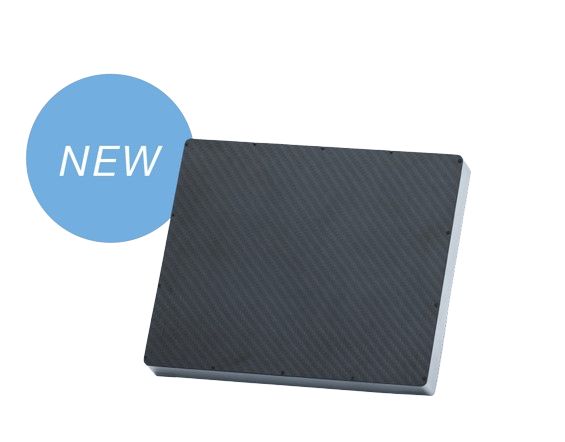
IGZO FPD
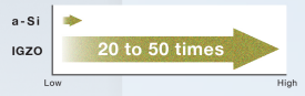
The IGZO semiconductor can conduct 20 to 50 times more current than the amorphous silicon (a-Si) semiconductor that was previously in wide usage, resulting in higher electron mobility and less leakage current. As a result, the amount of transmission per pixel can be increased, allowing for higher resolution image generation and noise reduction.
Comparison of electric current in an IGZO and a-Si semiconductor
Comparison of leakage current in an IGZO and a-Si semiconductor
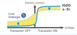
With IGZO, there is only a small leakage current when the transistor is turned off, and when the transistor is turned on, the electric current is strong. This allows for the generation of high-quality images with little noise.
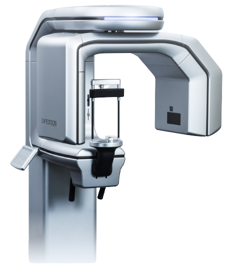
In Pursuit of High Image Quality
Superior image quality is produced by combining a high-quality X-ray generator, the key component for X-ray irradiation, with a variety of imaging technologies.
360° rotation
PreXion3D Expedition makes higher resolution images possible by obtaining information content from the entire circumference with 360° scanning with no image rendering necessary.
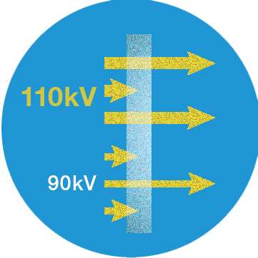
110kV High X-ray Tube Voltage
The high X-ray tube voltage of 110kV allows PreXion3D Expedition to deliver optimal image quality for all kinds of tissue while decreasing image artifacts.
16-bit grayscale
While many dental CT scanners have 14 bits (16,384 gradations), PreXion3D Expedition has a high gradation of 16 bits (65,536 gradations), enabling it to produce even smoother, higher resolution images.
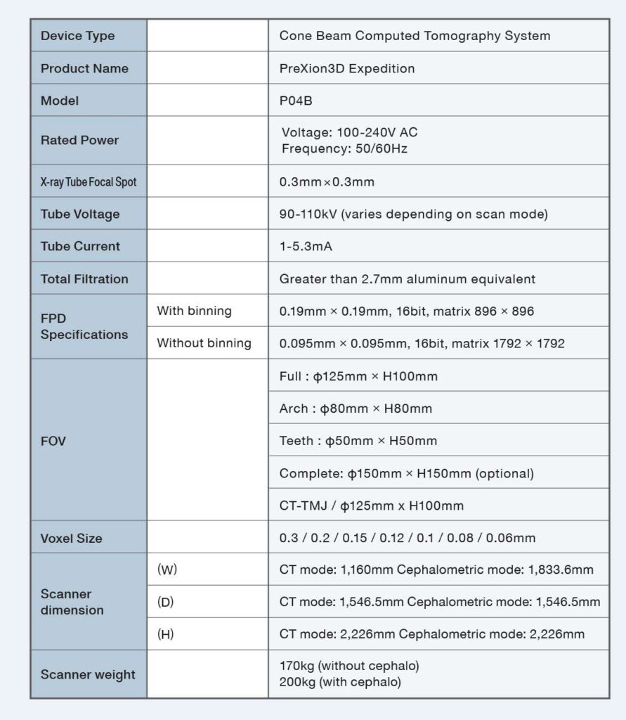
X-Ray Tube Focal Spot
0.3 x 0.3mm
The focal spot is the part of the X-ray tube where electrons strike a target and emit X-rays. PreXion3D Expedition features a 0.8mm X-ray tube focal spot, among the smallest in the industry.
Voxel Size
Minimum : 0.06mm
Maximum : 0.3mm
PreXion3D Expedition uses voxel sizes of 0.06 / 0.08/0.1/0.12/0.15/0.2/ 0.3mm. It displays 3D images with high resolution and high image quality. This is smaller than nearly every competitor.
Making Low Radiation Exposure a Reality
The PreXion3D Expedition is equipped with features such as pulse irradiation that emits X-rays intermittently and a low-dose mode, which can deliver high-image quality while minimizing radiation exposure, making more precise examinations possible.
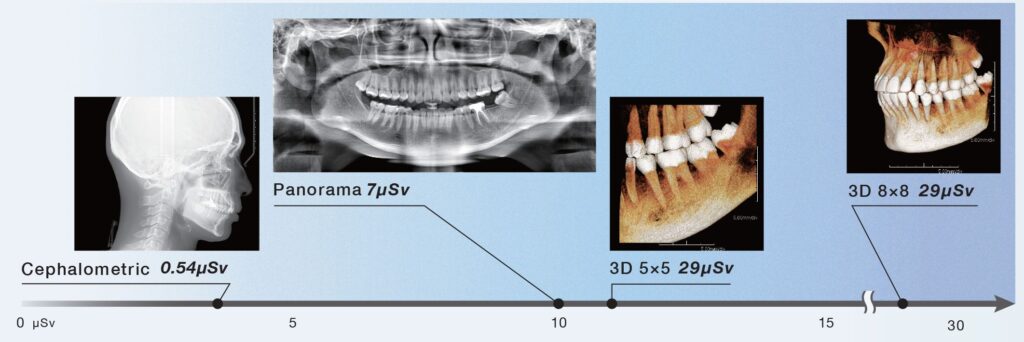
Exposure dose
Comparison of radiation dose when taking cephalometric, panoramic, or 3D scans. Although 3DCT provides a very large amount of information, it achieves low exposure.
Low Dose mode
Reduces radiation exposure and shortens scan time.

FOV |
Exposure dose |
Scan time |
|---|---|---|
5x5cm | 29uSv | 8 seconds |
8x8cm | 74pSv | 8 seconds |
12.5x10cm | 109uSv | 8 seconds |
Pulse irradiation
The exposure dose on the patient can be greatly reduced through the use of intermittent X-ray irradiation rather than continuous irradiation. This also reduces wear on the X-ray tube.

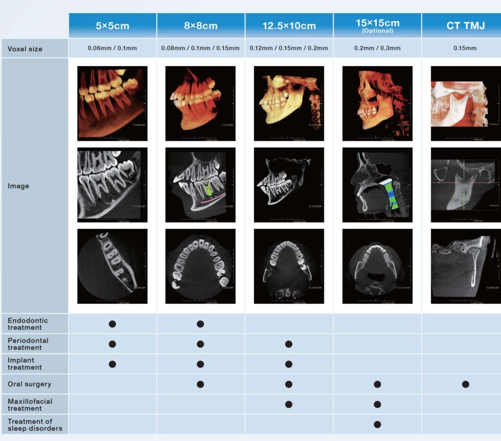
Fields of View (FOV)
PreXion3D Expedition is equipped with CT image acquisition capabilities applicable for all clinical situations at 5x5cm, 8x8cm, 12.5x10cm, and 15x15cm (optional), covering endodontic to full mouth treatment. PreXion3D Expedition uses a 360° rotation to generate high-resolution images for all FOVs.
2D One Shot Ceph
The optional cephalo unit for PreXion3D Expedition takes clear cephalometric images with one shot, reducing the patient scan time and exposure.
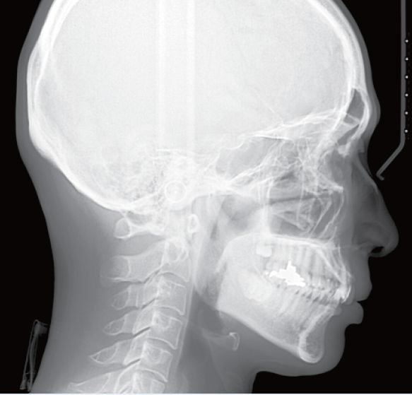
Ceph(LA)
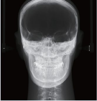
Ceph(PA)
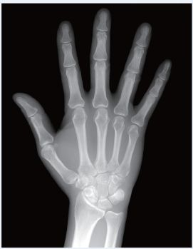
Carpus
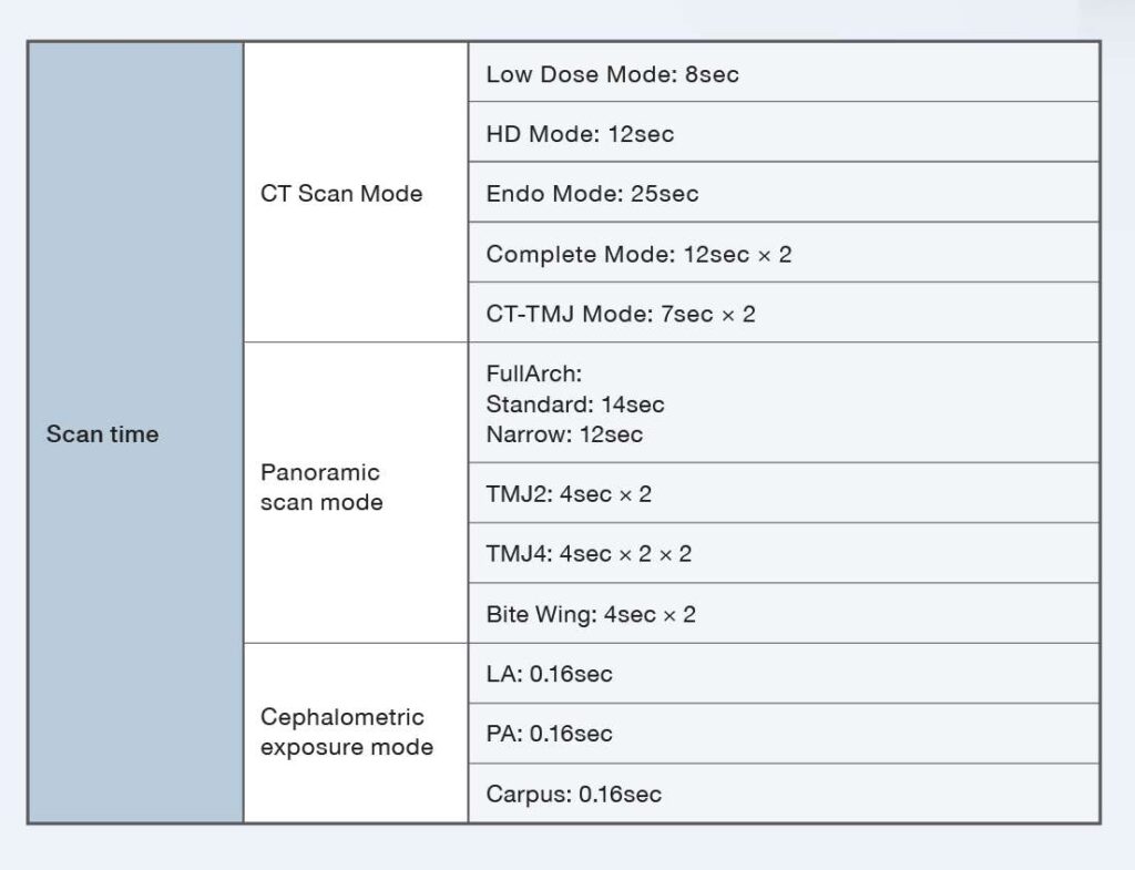
Scan Time |
CT Scan Made |
Low Dose Made: 8sec |
|---|---|---|
Scan Time | CT Scan Made | HD Mode: 12sec |
Scan Time | CT Scan Made | Endo Mode: 25sec |
Scan Time | CT Scan Made | Complete Mode: 12sec x 2 |
Scan Time | CT Scan Made | CT-TMJ Mode: 7sec x 2 |
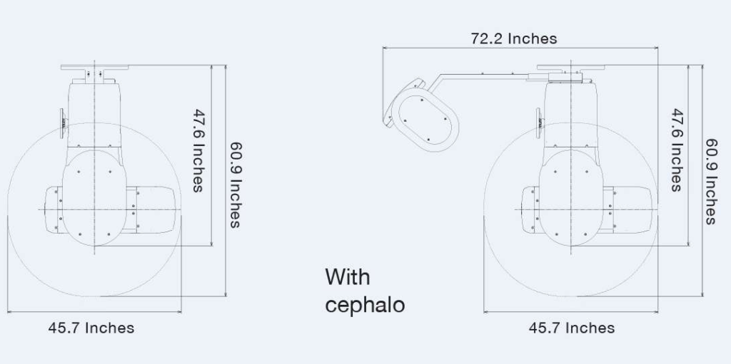
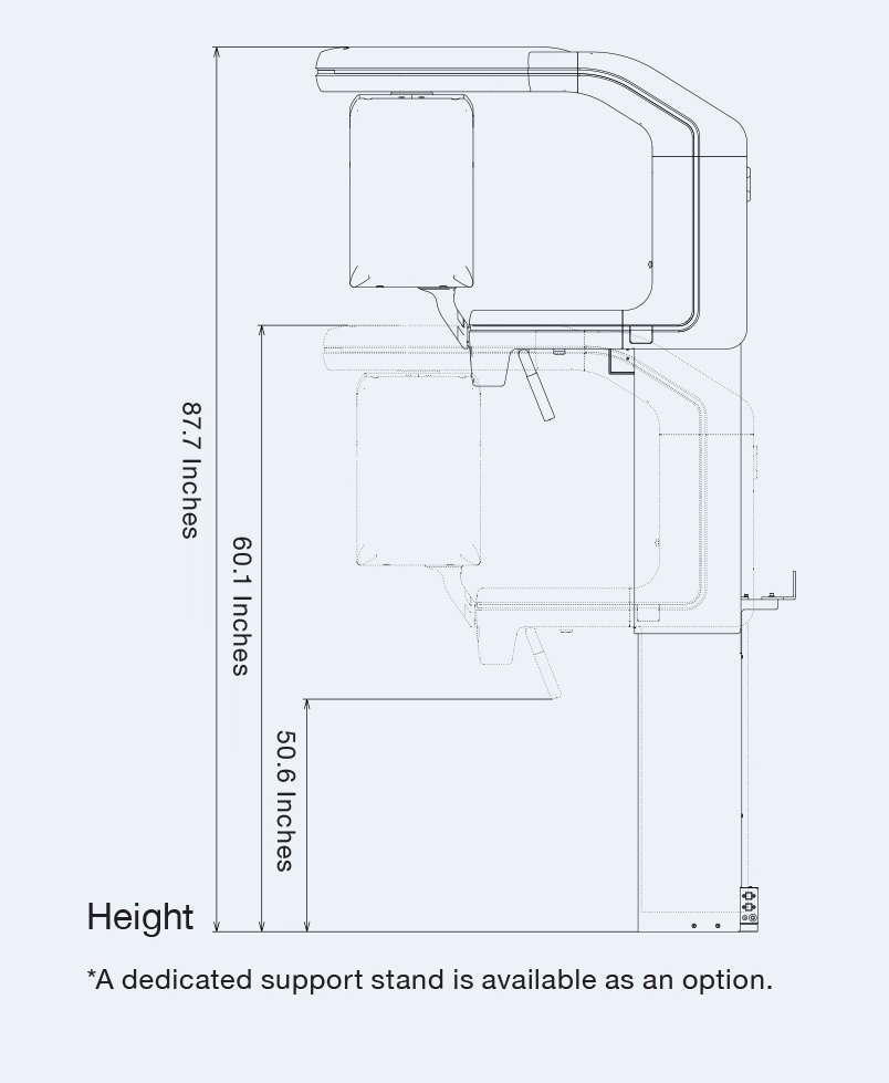
Dimensions